JPG 992 x 1248
JPG 2048 x 2176
JPG 580 x 990
AVI 280 x 495(70)
JPG 1161 x 1702
JPG 880 x 1070
JPG 1000 x 720
JPG 5120 x 6233
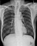
| JPG 992 x 1248 |
This digital chest radiograph was obtained with a posterior anterior (PA) projection using a 120 kVp radiographic technique. The image was recorded with a Kodak computed radiography (CR) system (Kodak CR900). The image was used in a research project to determine the best parameters to use for image processing enhancement. |
|---|---|
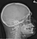
| JPG 2048 x 2176 |
A digital radiograph of the skull in a lateral projection illustrates how image processing may be used to equalize brightness in broad areas that would otherwise be over or underexposed. The processing is similar to the unsharp mask processing that is used to enhance digital photographs. The image was obtained from a Kodak CR900 system and processed using Enhanced Visualization Processing (EVP). |
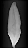
| JPG 580 x 990 AVI 280 x 495(70) |
Digital radiographs of this extracted tooth were obtained at 360 angles and 3D tomograms reconstructed. An individual view at full size is included in jpg format. A set of 70 images at 1/2 size illustrates the tooth as a rotating cine sequence (avi format). The data was is from an NIH sponsored project to develop a tooth atlas. |
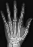
| JPG 1161 x 1702 |
A radiograph of the left hand taken with a solid state digital radiography detector (Selenium photoconductor, aSi-TFT readout, Kodak DR7100) demonstrates high bone detail. The direct recording of charge in these devices eliminates the detector blur present with indirect detector devices using optical scintillators. For this subject, a comlex fracture at the base of the fifth metacarpal is incompletely healed. |
| JPG 880 x 1070 |
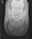
Dorsopalmar view of the equine foot on a laminitic horse. Notice that the gas dissecting up the dorsal hoof wall, the type VI solar margin fractures, and the Navicular bone can all be seen on this one view. Images were obtained using a Canon CXDI 50g digital radiography system supplied by Elkin. For more vetinary examples, see the Elkin Gallery or the Vetel Gallery. |
| JPG 1000 x 720 |
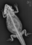
Full body radiograph of a lizard. Images were obtained using a Canon CXDI 50g digital radiography system supplied by Elkin. For more vetinary examples, see the Elkin Gallery or the Vetel Gallery. |
| JPG 5120 x 6233 |
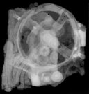
A two-thousand-year-old astronomical calculating device, the Antikythera Mechanism, radiographed at the National Archaeological Museum of Athens using an X-Tek 225 kV microfocus x-ray source and a Kodak ACR2000i CR system. The digital image pixels are 0.069 mm with 12 bit depth. For more information, see the X-tek web page or the Wikipedia page. |