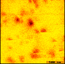
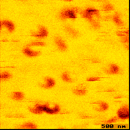
Figure 3. (a) False color fluorescence NSOM image of single DiIC12(3) molecules on a glass surface. (b) Single molecule fluorescence excited by linearly polarized light.
The electric field at the tip consists of a propagating field and a non-propagating evanescent field ringing the aperture (at the metal boundary). The ring like single molecule fluorescence patterns in Figure 3(b) result from excitation by the evanescent field but not from the propagating field which is polarized perpendicular to the molecules absorption dipole. Molecular orientation can be calculated from the shape of the image.