Near-Field Spectroscopy
To study single biomolecule dynamics we have developed near-field
spectroscopy. In near-field spectroscopy information is obtained
using spectroscopic techniques rather than imaging. The near-field
probe, placed over the molecule of interest, illuminates a sub-wavelength
area of the sample which enables spectroscopic studies of proteins in high
density in vivo or in situ situations. Detecting the emission
of a dye molecule or the intrinsic fluorescence of the protein of interest
can yield conformational information. The potential of near-field
spectroscopy lies in applying traditional far-field spectroscopic techniques
to the study of single biomolecules.
Initial experiments will focus on the problem of actin-myosin
binding. As a preliminary step towards this goal, we have imaged
polymerized actin labeled with TRITC phalloidin (Figure 4).
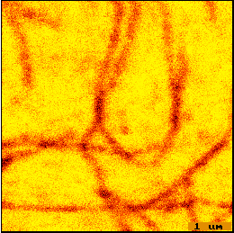
Figure 4. False color NSOM fluorescence image of TRITC phalloidin
labeled F-actin fixed to glass, cross linked with glutaraldehyde, and air
dried. Resolution is ~200 nm (limited by tip size).
Scanning in Solution