Post-Docs
Dr. Gurjit Mandair 
Gurjit Mandair obtained his PhD from the University of Southampton in 2004. Under the guidance of Dr. Andrea Russell he developed infrared mapping and imaging tools to characterize resin-bound combinatorial libraries and single-bead morphologies. In collaboration with Prof. Mark Bradley, he also synthesized anti-viral drug libraries [Tetrahedron Lett 2002] and screened them spectroscopically using a novel miniaturized flow cell attached to an infrared microscope. He joined the Morris group in 2005.
His early work, in collaboration with Karen Esmonde-White, involved using SERS to study hyaluronic acid in artificial synovial fluid standards, a potential biomarker for osteoarthritis. He then went on to use Raman spectroscopy to monitor mineralization events in osteogenic cell cultures. In collaboration with Dr. Ted Bateman (Clemson University), he used Raman to assess bone tissue composition from hindlimb suspended rodents in order to simulate the affects of short-term spaceflight on bone loss.
In a 5-year blinded study, he is currently testing the hypothesis of whether Raman spectroscopy can provide information on bone quality and whether it can distinguish between tissues that has undergone fracture and from tissues that has not (PI: Dr. Robert Recker, Creighton University). In parallel studies with Dr. Jason Long from the Goldstein Lab and Jessica Lopez (senior undergraduate), he is testing the hypothesis of whether bone composition as measured by Raman spectroscopy is commensurate with BMD as measured by micro-computed tomography. Such studies may expedite the diagnosis of fragility fractures and aid in the refinement of emerging heterogeneity-based bone quality metrics [CORR 2010].
 Dr. Peizhi Zhu
Dr. Zhu is currently working on the dynamics of mRNA and a translating ribosome using single molecule
imaging and microfluidic methods in collaboration with Nils G. Water lab, Chemistry and Mark Burns Lab,
Chemical Engineering. The overall goals of this project are to study the dynamics of ribosome movement along
mRNA, the penultimate step in protein assembly, and to progress towards a clearer understanding of the
processes that control this movement. A step-by-step approach was implemented to localize the ribosome
with near-codon (~2 nm) spatial resolution as it moves along mRNA. Dr. Zhu is also working on a bone
and mineral research program, which includes studies in decollagenated bone and bone structural
change under mechanical loads, in collaboration with Rams lab, Chemistry and David H. Kohn lab, School of
Dentistry.
Dr. Peizhi Zhu got his B.S. and M.S. from Nanjing University, and Ph.D. from University of Hong Kong. Before
joining the Morris Lab, he worked as a research associate at the Department of Molecular Biology at Scripps
Research Institute for about 2 years.
Dr. Paul I. Okagbare 
Dr. Okagbare received his Master of Science (M.Sc.) degree in analytical chemistry from the University of Ibadan, Nigeria in 2000. He worked with SmithKline Beecham as a Production Supervisor (2000-2001) and thereafter with GlaxoSmithKline as Manufacturing Manager (2001-2004).
He joined the doctorate program at Louisiana State University in the spring of 2005, where he did his PhD graduate research in the field of analytical and bioanalytical chemistry. His graduate research focused on developing polymer-based miniaturized (Lab-on-chip) tools and ultra-sensitive detection methods for high throughput biochemical analysis (high throughput screening in drug discovery) and single-molecule detection. These bio-modular microfluidic devices and wide-field imaging system provide for massive parallelization of fluidic processors and simultaneous read-out of assay results as demonstrated in a model-screening assay designed to monitor the activity of an enzyme, apurunic endonuclease 1. He got his PhD in May 2009 and received the 2009-2010 Outstanding Dissertation Award from Louisiana State University Graduate School . Dr. Okagbare joined Morris research group in 2009 as a Research Fellow, and his current research is focused on developing non-invasive Raman tomographic methodology for in-vivo evaluation of the time course of allograft osseointegration (bone graft incorporation and mineralization) in animal models. He is also investigating non-invasive methods for early diagnosis of bone-related diseases with potential utility in monitoring the efficacy of therapeutic agents as well as screening combinatorial libraries for specific targets.
Dr. Francis Esmonde-White
Dr. Esmonde-White completed his bachelor's degree in Chemistry at Concordia University in Montreal in 2002, with a specialization in computational chemistry. Following his undergraduate studies, he completed his Ph.D. studies at McGill University in Montreal in 2008. His doctoral studies included the development of a portable photon time-of-flight instrument and annular imaging instrument for measuring tissue optical properties and data analysis methods to correct for light scattering in biomedical spectroscopy.
He joined the Morris group in 2008, and has worked on a number of projects related to transcutaneous Raman spectroscopy and tomography. In particular, he has developed methods taking into account sample geometry, the optical properties (Raman, absorption and scattering), and the effects of multiple sample layers. Among other projects, he has developed: a new method for creating multilayer geometrically-accurate tissue phantoms, software methods for correcting Raman imaging measurements and recovering spectra from individual fibers in fiber bundles, a new method for predicting optical sampling volumes using the NIRFAST diffuse optical tomography software, and new conformal fiber optic probes for Raman tomography. His work in the Morris lab has included studies of ex-vivo human and animal tissues for non-invasive diagnostics of musculoskeletal tissues.
Dr. Karen Esmonde-White 
If one can see “the world in a grain of sand”, imagine how much chemical information is in a drop of fluid. Karen Esmonde-White is working on developing Raman spectroscopy-based techniques to identify joint damage associated with early-stage osteoarthritis (OA). It is my goal to develop a method that will diagnose OA before a patient even feels OA-related pain! How am I doing this? I hypothesize damage associated with OA that is expressed in the fluid surrounding the joint, called synovial fluid, can be observed with Raman spectroscopy. These changes in the chemistry and/or mechanical properties (such as viscosity) of synovial fluid have been proposed as markers of early-stage OA detection.
For synovial fluid analysis, I am collecting Raman spectra of fluid drop deposition (also known as the “coffee-ring effect”) to examine subtle changes in composition that may be an early indication of joint damage. In the Morris lab, we are collaborating with colleagues in the UM Medical School in a pilot study to test this hypothesis. Early results are promising! The deposition pattern, as seen by microscope photographs, together with Raman spectra of dried synovial fluid drops can be used as OA markers. Along the way, I have found some interesting chemistry relating to the drop deposition process and how it affects collection of Raman spectra from these dried drops. Some early work in the drop deposition project was also done by Gurjit Mandair, a post-doc in our lab.
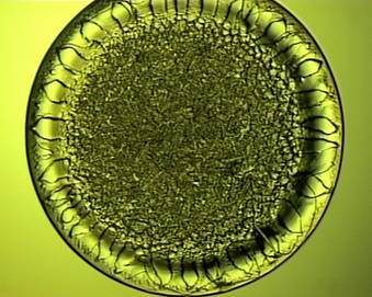
The figure above shows what a dried synovial fluid drop looks like under our microscope. Because the microscope is configured for either light microscopy or Raman microspectroscopy, we can also collect near-infrared Raman spectra. The shape of the drop is round, although the shape varies slightly depending on the viscosity of the fluid and the surface chemistry of the microscope slide. The drop itself has very interesting features: there is a thin-film ring on the outer edges of the drop and fern-shaped crystals in the center of the drop.
 Dr. Katherine Cilwa
Katherine Cilwa received her PhD in Physical Chemistry from the Ohio State University in 2011. At Ohio State Katherine experimentally and theoretically characterized plasmonic mesh systems active in the infrared. By modeling the interaction of simultaneously occurring plasmonic states she demonstrated the mixed boson/fermion-like behavior of surface plasmon-polaritons.
She also designed a non-destructive method of collection and component analysis of wavelength-scale particulate matter by placing 3-5 µm dust particles each within their own channel of plasmonic mesh and using FTIR imaging microscopy to obtain scatter free absorption spectra of 63 individual particles [J. Phys. Chem. C 2011]. This work included modeling infrared scattering and absorption spectra of multi-component individual dust particles using dielectric medium theories, through which she identified key issues quantifying component concentration in individual dust particles and demonstrated deviation of particulate materials from bulk absorption behavior. Katherine joined the Morris group in 2011. Her work focuses on examining the effects of aging in mice on bone material properties using nanoindentation and Raman spectroscopy in collaboration with Dr. Ken Kozloff (Department of Orthopaedic Surgery), Dr. Bernard Halloran (UCSF Department of Medicine), and Amgen.
Dr. Alexander Khmaladze 
Dr. Khmaladze received his Ph.D. in Applied Physics from the University of South Florida in 2008. He is currently working on analyzing the quality of ex vivo produced oral mucosa equivalent (EVPOME) constructs by Raman spectroscopy in collaboration with Dr. Stephen Feinberg from the Department of Surgery at the University of Michigan.
Additionally, his research interests also include non-linear scanning microscope design, hyperspectral imaging of live Oocytes via Coherent Anti-Stocks Raman Scattering (CARS) Microscopy and three-dimensional digital holographic cell imaging. He is the author of over 30 peer-reviewed papers and conference proceedings.
 Dr. Mekhala Raghavan
Dr. Raghavan completed her bachelor's degree in Instrumentation & Control Engineering at Anna University in Chennai, India in 2005. She received her PhD in Biomedical Engineering from the University of Michigan in 2010. Her doctoral and post-doctoral work advances the understanding of bone quality using Raman spectroscopy along multiple directions: investigation of age-dependent bone quality, analysis of microdamage in diseased bone, BMP signaling during bone development, quantification of molecular disorder in brittle bone disease, and analysis of mineral deformation behavior. She is also a postdoctoral associate at the Center for Global Health, University of Michigan.
Dr. Bo Gong
details coming soon...